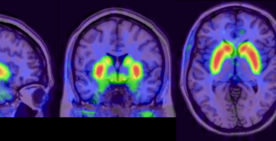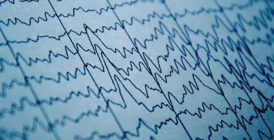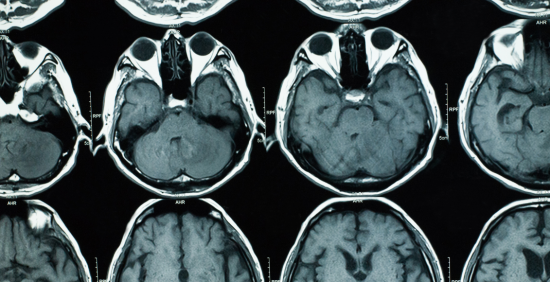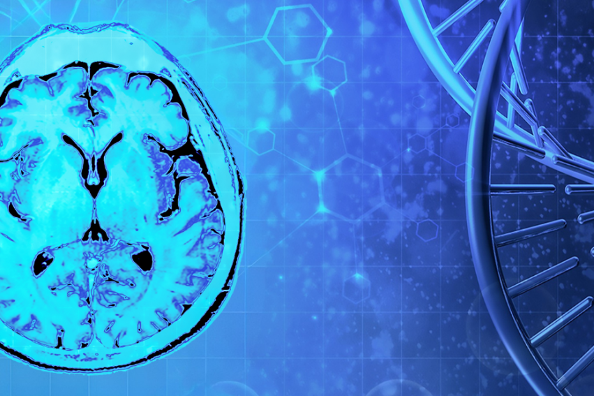


Neuroimaging comprises techniques such as magnetic resonance imaging (MRI), Positron emission tomography (PET) and electroencephalography (EEG to study the brain).
We investigate brain structure, function, physiology and metabolism across the breadth of psychiatric and neurological disorders.
Our overall aim is the continued development and implementation of neuroimaging for:
- better diagnosis,
- improved understanding of the biological mechanisms behind disorders,
- enhanced prediction of which patients respond differently
- and clearer grouping of patients for translational and clinical studies.
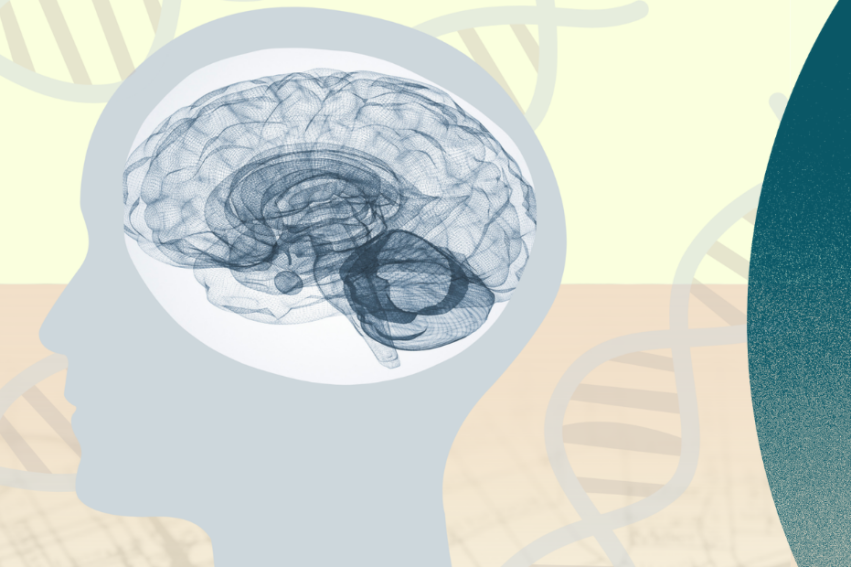
Through our technological innovations, we are extending our palette of measurable biological markers to include those were previously inaccessible; for example, studying the brain’s “drainage system” which is known as ‘glymphatics’.
We develop novel image acquisition and analysis techniques to improve access to brain imaging techniques and to visualise previously unattainable brain function. In doing this, we have created tools and infrastructure to support continued access to our neuroimaging data for the open science community.
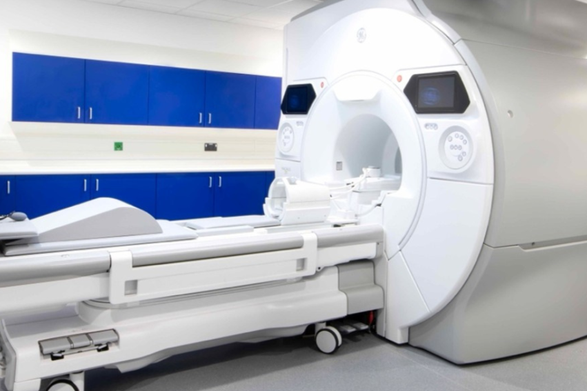
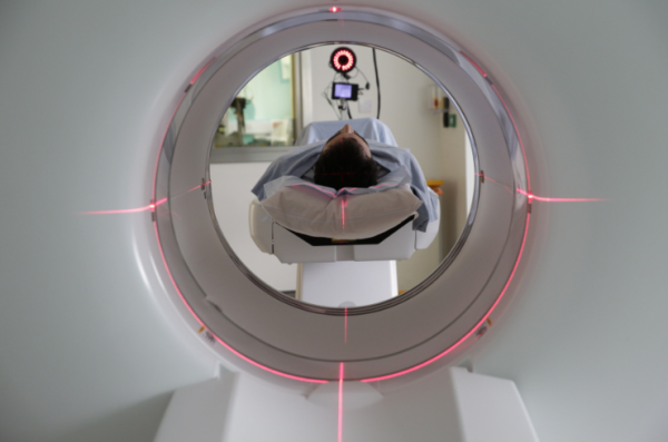
By advancing techniques such as silent MRI and portable MRI, we are improving accessibility, reducing costs, and enhancing the scanning environment for some of our most unwell and under-represented patients.