Anaemia in pregnancy linked to differences in babies' brain development
Child Mental Health and Neurodevelopmental DisordersNeuroimaging
Babies born to mothers who were anaemic during pregnancy have smaller brain volumes than babies born to mothers who were not, according to a new study by researchers at the Institute of Psychiatry, Psychology & Neuroscience (IoPPN) and the University of Cape Town.
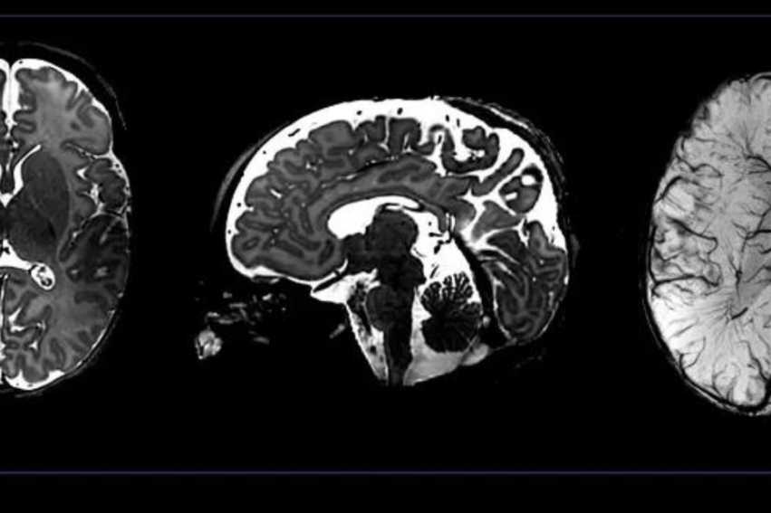
Using a portable MRI scanner, the research found that measurable differences in brain volumes emerge from around 12 months of age, offering some of the earliest evidence yet that maternal anaemia shapes infant brain development in the first year of life and beyond.
The study, published in Brain Communications, followed 394 mother-infant pairs in Cape Town, scanning a subgroup of the infants’ brains at several points between three months and two years old. Babies born to mothers who had been anaemic during pregnancy had, on average, around 4% smaller total brain volume than babies born to non-anaemic mothers. Differences were also found in three specific regions: the putamen, the caudate nucleus, and the corpus callosum – structures involved in movement, learning, emotion regulation, executive functioning, and communication between the two halves of the brain.
Over the first two years of life, the gap widened. The corpus callosum was around 4% smaller in the anaemia-exposed group by 12 months, but by 24 months, that gap had grown to 6%. This builds on previous research by Ringshaw, Donald and colleagues on toddlers and school-age children in South Africa, that found comparable brain volume differences (between 4-8%) in these same brain regions. The new study shows that these differences can be detected as early as one year of age.
The observed differences in regional brain volumes were still evident despite most of the mothers in this cohort only having mild anaemia. The researchers say these findings identify antenatal maternal anaemia as a strong risk factor for altered child brain structure and highlight the importance of preventing and treating anaemia before and during pregnancy.
Seeing such prominent volumetric differences in infants exposed to anaemia in the womb reinforces the importance of optimising interventions for women both before and during pregnancy.
Jessica RingshawFirst author and researcher at King's IoPPN and the University of Cape Town
Jessica Ringshaw, First author and researcher at King’s IoPPN and the University of Cape Town said: “What is striking about these findings is how early we are able to detect them. Seeing such prominent volumetric differences in infants exposed to anaemia in the womb reinforces the importance of optimising interventions for women both before and during pregnancy.
In settings with multiple overlapping risk factors such as malnutrition and infectious disease, it is important to think about how they may be working together to drive anaemia and, in turn, impacting brain development. By tailoring interventions to the local context, we can make a meaningful contribution to society by giving children the best chance of reaching their full developmental potential—not only surviving, but thriving.”
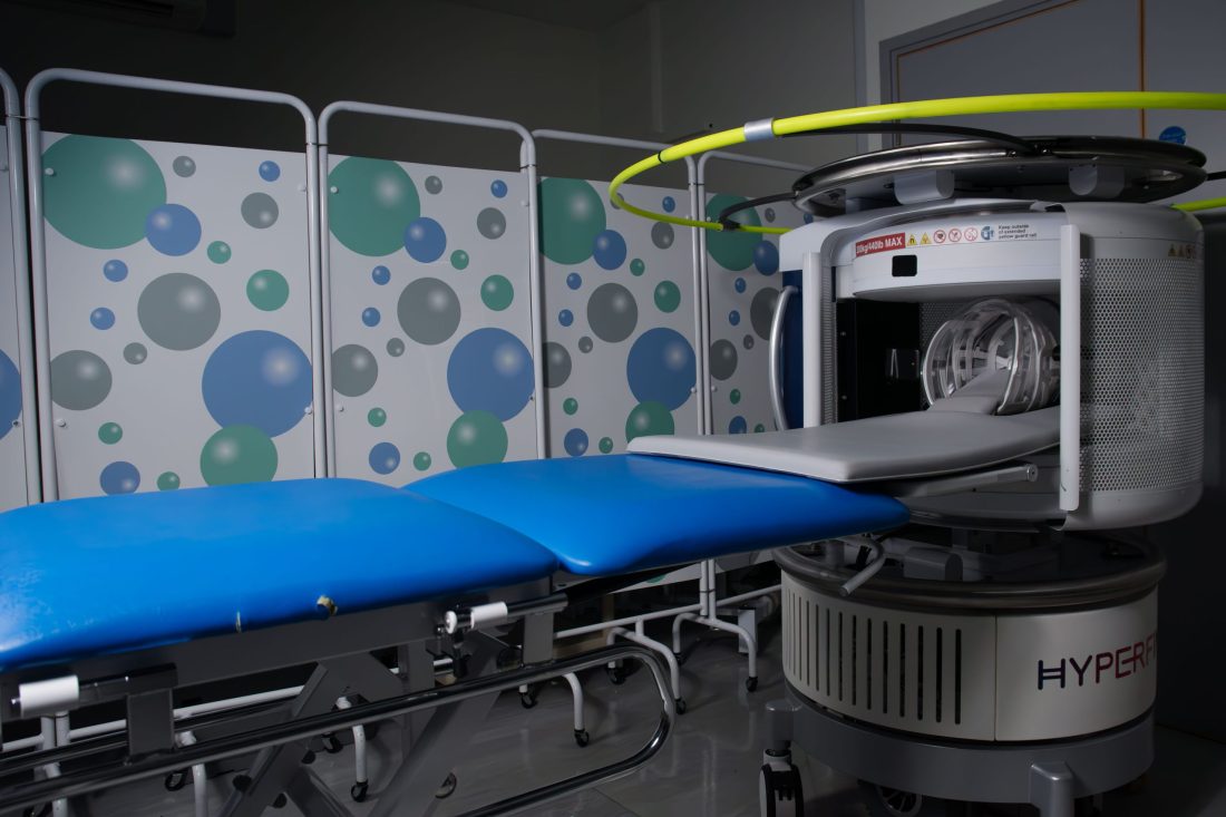
A portable low-cost, ultra-low-field, energy-efficient MRI scanner was used by the researchers alongside a regular high-field scanner.
The study is also the first to test whether a portable, low-cost, energy-efficient MRI scanner can pick up these differences as reliably as a conventional hospital scanner. The team used both a standard 3 Tesla scanner and a lightweight 64 millitesla device that can be brought directly to communities, rather than requiring families to travel to a hospital. It also requires less infrastructure and specialised expertise, and is significantly quieter than high-field scanners, meaning sleeping infants can easily be scanned without sedation.
The researchers say this opens the door to similar studies, including research assessing the efficacy of interventions, in parts of the world where conventional scanners are scarce.
This study has proven that our novel technology can measure the impact of malnutrition on the developing brain, at scale, in some of the most challenging environments. This will provide an early, sensitive assessment of planned interventions.
Professor Steve WilliamsProfessor of Neuroimaging at King’s College London (part-funded by NIHR BRC: Maudsley)
Anaemia in pregnancy affects more than a third of pregnant women worldwide, with the highest rates in sub-Saharan Africa and South Asia. While anaemia is most often driven by iron deficiency due to diet or the body not being equipped to absorb it properly, infections such as HIV can also cause anaemia. The team found that mothers in the study living with HIV had a greater risk of being anaemic in pregnancy, which the researchers say may be due to indirect effects of inflammation on how the body process iron from the food you eat.
Professor Kirsten Donald, senior author and Principal Investigator of the Khula South Africa cohort at the University of Cape Town, said:“Anaemia in pregnancy, much of it related to iron deficiency, affects millions of women and children worldwide. Until now, our ability to see what this means for early brain development has been very limited in the settings carrying the greatest burden. Measuring these effects directly opens up an important new avenue for understanding which maternal nutrition interventions can make a meaningful difference to children’s developmental trajectories.”
This research was funded by the Wellcome Trust, Wellcome LEAP, the Gates Foundation, and the Science for Africa Foundation. This research forms part of the projects from the UNITY network, funded by the Bill & Melinda Gates Foundation. Professor Steve Williams is supported by the NIHR BRC: Maudsley.