
The neuroimaging suite is situated at the rear of the ground floor. It comprises a new 3T functional GE MRI (fMRI) scanner, a control room, an image analysis room, clinical preparation rooms and a research office.
The coexistence of MRI, EEG, TMS, the Virtual Reality Suite, behavioural assessment and biological sampling facilities under the same roof provides a unique environment for multimodal imaging and integrated collection of large data sets. This imaging facility is perfectly suited for the assessment of less ambulatory patients, especially when serial assessments are needed during the disease course: for example, after stroke, surgery or brain injury.
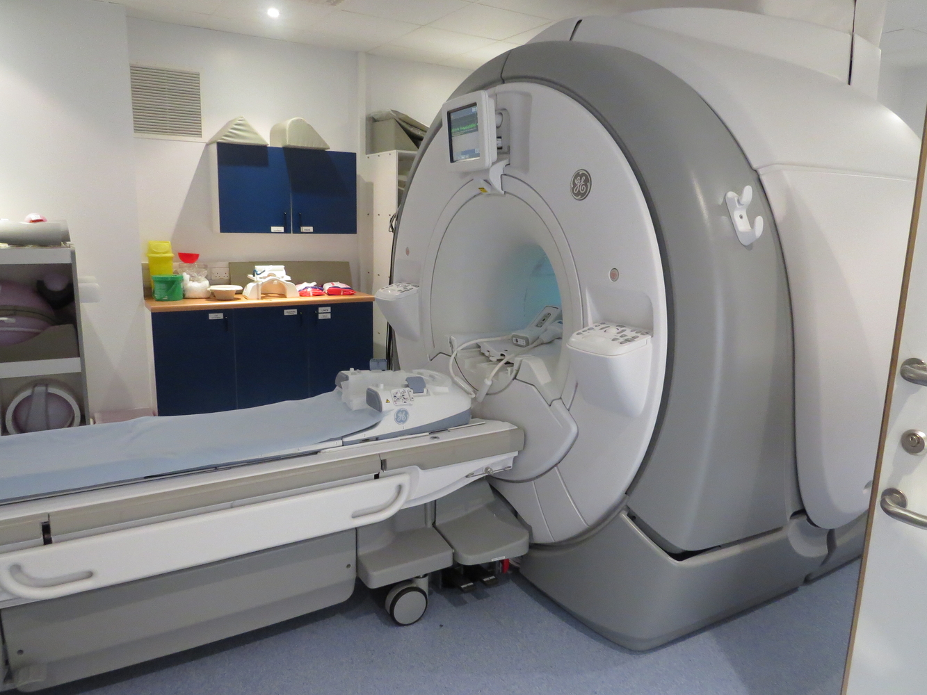
Scanner:
- A 3Tesla GE Healthcare MR750 MRI scanner was commissioned and installed in the summer of 2011, and was specified to provide state-of-the-art MR imaging modalities in all areas of clinical research. For this purpose, it has been equipped with RF coils for neuro (brain, cerebro-vascular and spine), abdominal and extremity imaging.
- In addition, the scanner also includes capabilities for multi-nuclear spectroscopy (1H, 13C and 31P).
- The primary clinical target of this unit is the application of MRI to the study of acute and chronic presentations of traumatic brain injury (head trauma, stroke and tumour). To facilitate this, the unit is also equipped with all devices necessary to monitor and maintain anaesthetised patients while scanning. Neuro-applications are a primary research objective of this unit; to this aim the scanner has capability for high-speed, high-resolution Functional Imaging, Diffusion Tensor Imaging, MR Angiography and Arterial Spin Labelling.
- Another target area for this unit is the application of MR imaging technology to the characterisation of candidate psychoactive compounds
Clinical Preparation Area:
- With its own dedicated room, it is equipped with all facilities necessary for clinical assessment of patients and healthy control subjects prior to and after scanning. The room has access to the MRI scanning suite via a dedicated RF screened hatch, incorporated into the room’s RF shield, in order to collect blood samples from subjects whilst being scanned. The room also houses a high-speed centrifuge and a -20C freezer for short-term storage of serum samples for subsequent analysis.
EEG
- Two state of the art electroencephalography (EEG) purpose built rooms for both supine and sitting recording and use with computer stimuli.
- Electronic and acoustic shielding provides a uniquely optimal environment for the study of subjects undergoing EEG monitoring; and allowing highest-quality multi-modal data collection.
Transcranial Magnetic Stimulation (TMS)
- The CRF is able to host TMS research studies (especially in conjunction with EEG) and now have ‘Visor2' which accurately evaluates functional organisation of the human cortex using non-invasive, navigated transcranial magnetic stimulation.
Interview and image processing rooms
- Adjacent to the unit there is a room for interviewing, screening and training of subjects before and/or after scanning. A dedicated team is in charge of implementing software programs for administration of sensory and cognitive paradigms for functional imaging. A very large range of hand held devices (button boxes, joysticks, pressure sensitive controls, etc.), have been built for the execution of such functional paradigms in the scanner.
Access & Contact:
- Please contact the Clinical Research Facility Manager, Elka Giemza to discuss undertaking your project at the CRF or Maryla Kaminska (mri.booking@kcl.ac.uk) for scanning-related research queries.

